Breast cancer screening: the mammography equipment of today and tomorrow
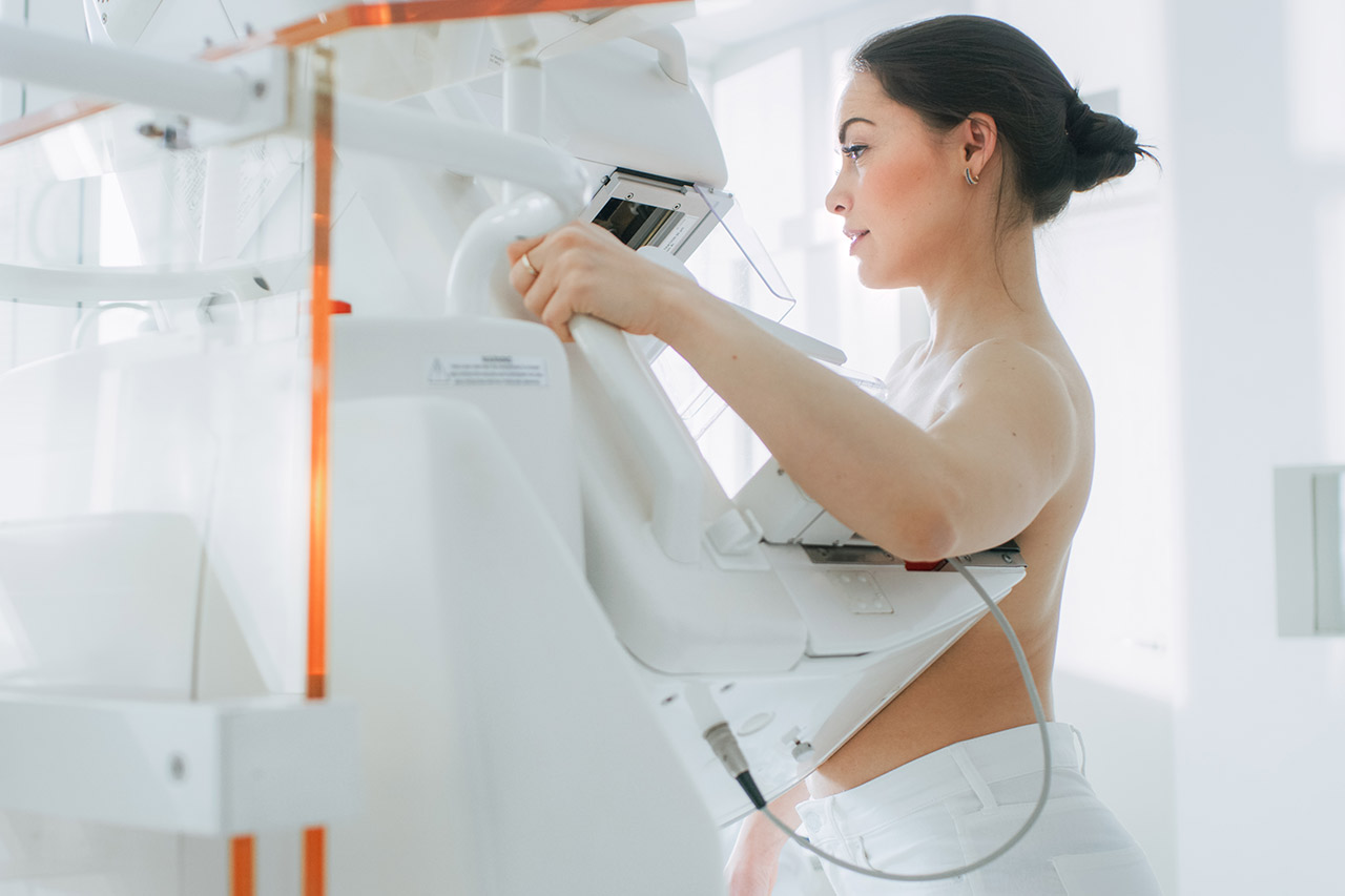
Screening for breast cancer is a major public health issue. With more than 2 million new cases worldwide each year, breast cancer is the leading cause of cancer death for women. However, early detection drastically reduces its consequences. Currently, in most European countries, screening is carried out every 2 years, for all women aged 50 to 74 without any apparent symptoms or particular risk factor. It is performed with a 2D digital mammogram, and potentially an ultrasound. The detection of an anomaly leads to additional examinations (ultrasound, MRI, tomosynthesis) followed by a biopsy, and possible treatment. Therefore, it is crucial to have more reliable, faster and less intrusive screening techniques in order to better diagnose women and men (approximately 1% of people affected by breast cancer are men) and reduce the impact of breast cancer on their health.
In recent years, breast cancer screening techniques have evolved and improved significantly. Alcimed is looking at the latest trends aimed at improving patient diagnosis.
Tomosynthesis, the start of a new era for breast cancer screening, or an innovation that is still too flawed?
Several breakthroughs in breast cancer screening techniques have occurred in recent years. First, the authorization of digital mammography in 2008 as part of organized screening in France, was used alongside analog mammography, then replaced it in most European countries. More recently, tomosynthesis has appeared.
First marketed by Hologic in 2011, tomosynthesis enables imaging “between 2D and 3D”. It has undoubtedly increased the detection of tumors. While perceived as a great advance, the improved performance of lesion detection has led to an overdiagnosis, and an increased number of biopsies, limiting the desire for dissemination of this type of equipment. This mammography technique also results in more radiation (depending on the device) and doubles the time to read the results. The generalization of tomosynthesis for screening is far from certain. Although the United States has recommended its systematic use in screening, the HAS in France has not validated it as of today.
The mammography of tomorrow: promising technologies for improving diagnostics
Other advances to improve diagnosis have also emerged in recent years, such as angiomammography. This technique combines an iodinated contrast agent with mammography and presents a diagnostic performance significantly superior to digital mammography.
To make the diagnosis more reliable, work is underway and aims to create a 3D mammography system that would operate by phase contrast. This solution has the advantage of no longer requiring breast compression like current solutions. It would also make it possible to have a complete 3D view (tomography), to detect microcalcifications still undetectable to date, and to greatly increase the detection of tumors in dense breasts. While product development, testing and marketing times are relatively long, this type of solution could be available and used within fifteen years.
Artificial intelligence: an impact on all aspects of mammography
Beyond the evolution of imaging techniques, artificial intelligence will become increasingly important, from the organization of screening to the analysis of results.
Artificial intelligence is very likely to impact breast cancer screening by allowing a personalized assessment of the risk of developing breast cancer. Projects like MyPeBs (My Personal Breast Screening, an international project funded by Unicancer) shows the importance of the trend towards customizable screening. This type of screening would make it possible to better diagnose women most at risk. The interval between two screenings could be determined according to many criteria (which remain to be defined) such as family history, breast and tissue structure, breast density, age, or body mass index.
In terms of the evolution of screening equipment, it is at the software level where a big changes are taking place. Artificial Intelligence is playing an increasingly important role in helping physicians read and analyze results. CAD (Computer Aided Diagnosis) is already in use and promises to become more and more efficient with the enrichment of radiomics databases.
The major challenge today is to develop more efficient equipment, which reduces false negatives without increasing false positives to avoid unnecessary biopsies. Tomosynthesis has certainly made it possible to reduce false negatives, but still leads to too much overdiagnosis. Projects of new techniques are moving in this direction, and by taking advantage of the contribution of artificial intelligence, should result in a marked improvement in diagnostics. We are convinced that the potential of these solutions could ultimately reduce the mortality linked to breast cancer today.
Interested in this subject? Discover our position paper on INNOVATION IN ONCOLOGY and our achievements in technological state of the art.
About the author
Julia, Consultant in Alcimed’s Healthcare team in France
Do you have an exploration project?
Our explorers are ready to discuss it with you